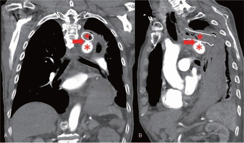
Figure 2.

Computed tomographic angiography visualized an aortoesophageal fistula with an ulcer-like projection (red arrow head) on the aortic arch (red asterisk) where the esophageal stent (red star) touched. (A) Coronal position. (B) Sagittal position.
