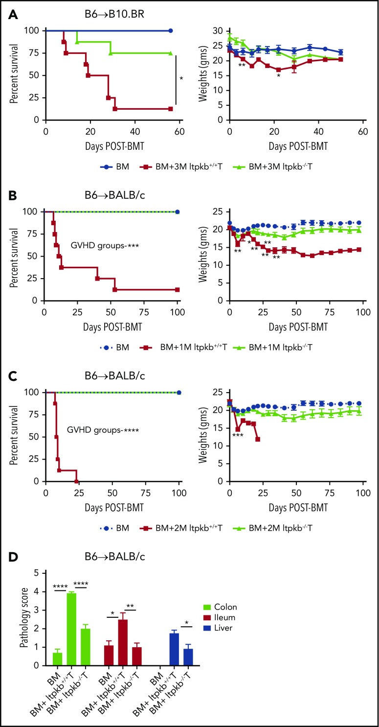
Figure 1.
Induced Itpkb loss in donor T cells prevents their capacity for aGVHD lethality. (A) Survival and weight curves are shown for irradiated B10.BR recipients that were given B6 BM (107), with or without B6 purified T cells (3 M is 3 × 10⁶ cells) from Itpkb+/+ donors or tamoxifen-treated donors to delete Itpkb (Itpkb−/−), as described in “Methods.” (B-C) BALB/c recipients were lethally irradiated on day −1 and infused with B6 BM (107), with or without B6 Itpkb+/+ or Itpkb−/− purified T cells on day 0 (panel B, 1 M is 1 × 10⁶; panel C, 2 M is 2 × 10⁶ cells). Survival and weight curves are shown (n = 5 mice/BM group; n = 8 mice/BM+T group). (D) Histopathology scores of tissues (liver, ileum, and colon), from BALB/c recipients of transplanted B6 BM (107), with or without B6 Itpkb+/+ or Itpkb−/− purified T cells (1.5 × 10⁶). Tissues were harvested on day 7 after transplantation, stained with hematoxylin and eosin, and scored for GVHD severity according to a semiquantitative scoring system (0-4, with 4 denoting more severe disease). Mean ± standard error of the mean (SEM); n = 5 to 6 mice per group. One experiment was performed. *P < .05, **P < .01, ***P < .001, and ****P < .0001.

