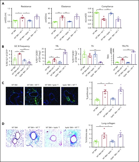
Figure 5.
Donor T-cell Itpkb expression is critical for GC reactions and cGVHD in the BO model. B10.BR mice were given cyclophosphamide (120 mg/kg per dose, days −3 and −2) and underwent TBI (8.3 Gy, day −1) followed by day 0 infusion with B6 WT or Itpkb−/− T-cell–depleted BM, with or without purified WT or Itpkb−/− donor T cells (70 × 103). (A) Pulmonary function was evaluated at 8 weeks after transplantation. (B) Recipient splenocytes were harvested at 6 to 8 weeks after transplantation and stained with fluorophores to quantify GC B cells (CD19+ Fas+ GL7+), Tfh cells (CD4+Foxp3-CXCR5+PD1hi), and Tfr (CD4+Foxp3+CXCR5+PD1hi) cells, and the Tfh/Tfr ratio was calculated. (C) Representative lung immunoglobulin deposition images and quantification. Confocal images were acquired on an Olympus FluoView500 Confocal Laser Scanning Microscope at original magnification ×200. (D) Collagen deposition in the lung was assessed by trichrome staining that identifies collagen in blue. The percentage of collagen deposition area was quantified by Fiji software. Four to 5 mice were analyzed for each group in each assay. Results shown are representative of 2 independent experiments with similar results. Data are shown as the mean ± SEM. *P < .05, **P < .01, and ***P < .001.

