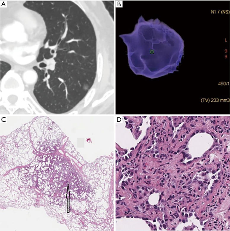
Figure 1.
Minimally invasive adenocarcinoma (MIA) in a 62-year-old woman. (A) The axial CT image reveals a pGGN with the diameter of 9.7 mm × 8.9 mm and with slight lobulation and adhesion to the pleura in left lower lobe; (B) the three-dimensional volume of the pGGN was 233 mm3, then we calculated that its mass was 80 mg; (C) low-magnification of the histologic specimen (Hematoxylin Eosin, 40×) shows darkly stained region (black arrow); (D) high-magnification of the histologic specimen (Hematoxylin Eosin, 200×) shows a small amount of tumor cells infiltrate the lung interstitial tissue with invasion thickness ≤5 mm. CT, computed tomography; pGGN, pure ground-glass nodule.

