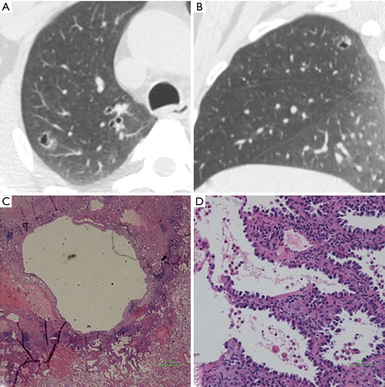
Figure 5.
GGN with cystic airspace in a 53-year-old woman. (A) Axial and (B) sagittal CT image show a thin-walled cavitary GGN with the diameter of 13.7 mm × 11.3 mm in right upper lob. (C) Low-magnification (Hematoxylin Eosin, 40×) and (D) high-magnification (Hematoxylin Eosin, 200×) of the histologic specimen show an irregular cystic cavity and tumor cells grow along the pre-existing alveolar wall. GGN, ground-glass nodule; CT, computed tomography.

