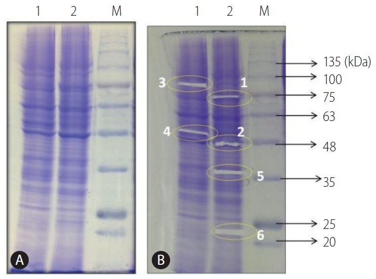
Figure 2.

(A) Protein profile of HepG2 (P) and (R) cells on 10% SDS-PAGE stained with Coomassie brilliant blue. (B) Excision of protein bands from HepG2 cells [lanes: 1-HepG2 (P); 2-HepG2 (R); M-BLUelf Prestained Protein Ladder]. HepG2 (P), HepG2 parental; HepG2 (R), HepG2 sorafenib-resistant; SDS-PAGE, sodium dodecyl sulfate polyacrylamide gel electrophoresis.
