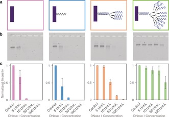
Figure 3.

Structural stability test against nuclease digestion. a) DNA brick nanostructures with four different surface densities were studied: bare (pink), single (blue), triple (orange), and nonuple (green). All assembled DNA brick nanostructures were incubated at 37 °C for 1 hour with varying DNase I concentrations (0 to 100 U mL−1). b) Incubated samples were characterized via gel electrophoresis (Lane 1: control; Lane 2: 5 U/L of DNAse 1; Lane 3: 0 U/L of DNAse 1; Lane 4: 50 U/L of DNAse 1; Lane 5: 100 U/L of DNAse 1). c) Gel band intensities from each gel were quantified via a gel‐imaging software, then normalized such that the intensity of the control is always 1. Data from three separate gel results were averaged, and standard deviations were calculated..
