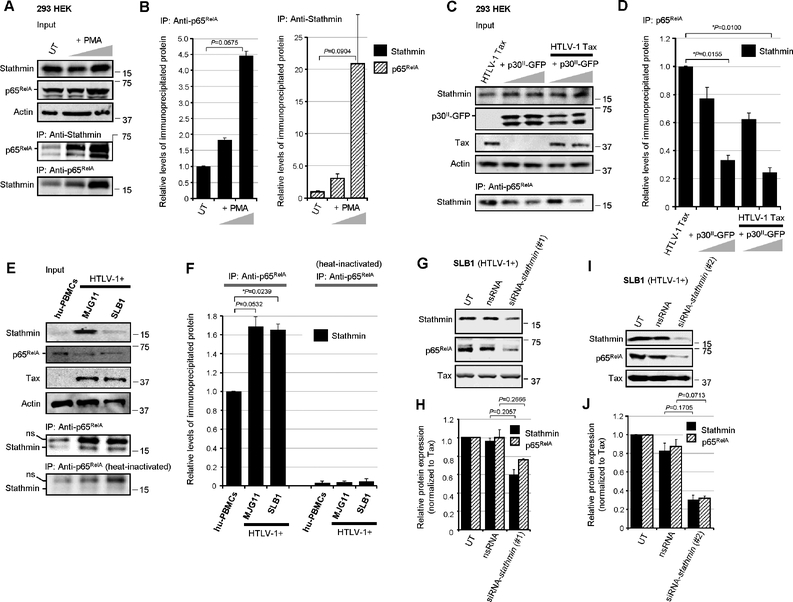
Fig. 3.
Stathmin interacts with the NF-κB p65RelA subunit in HTLV-1-transformed ATLL cells. (A) p65RelA-Stathmin protein complexes were immunoprecipitated from precleared whole-cell lysates prepared from PMA-stimulated 293 HEK cells using Protein-G-agarose (20 ml of a 50% slurry) and monoclonal Anti-p65RelA and rabbit polyclonal Anti-Stathmin/Op-18 antibodies. The relative input levels of Stathmin, p65RelA, and Actin are shown in the upper panels. The precipitated bound p65RelA and Stathmin proteins were resolved by SDS-PAGE and detected by immunoblotting (lower panels). UT, untreated cells. (B) The relative levels of the immunoprecipitated Stathmin (left) and p65RelA (right) proteins in A were quantified by densitometry analysis of the immunoblot bands. (C) The formation of HTLV-1 Tax-induced p65RelA-Stathmin immune-complexes is markedly diminished in cotransfected cells expressing increasing amounts (2.5 and 5.0 mg) of p30II-GFP. The relative input levels of HTLV-1 Tax, p30II-GFP, Stathmin, and Actin are shown (upper panels). The Stathmin/Op-18 protein, complexed with p65RelA, was co-immunoprecipitated using Protein-G-agarose and an Anti-p65RelA antibody and detected by immunoblotting (lower panel). (D) The relative levels of the immunoprecipitated Stathmin protein in C was quantified by densitometry. (E) p65RelA-Stathmin protein complexes were immunoprecipitated from precleared extracts prepared from cultured/activated hu-PBMCs and the HTLV-1-transformed ATLL T-cell-lines, MJG11 and SLB1, using a monoclonal Anti-p65RelA antibody. As a negative control, the Anti-p65RelA antibody was heat-inactivated prior to its use in immunoprecipitation reactions. The relative input levels of Stathmin, p65RelA, Tax, and Actin are shown in the upper panels. The precipitated Stathmin protein was resolved by SDS-PAGE and detected by immunoblotting using a rabbit polyclonal Anti-Stathmin/Op-18 antibody (lower panels). A non-specific band (ns) is also indicated. (F) The relative levels of immunoprecipitated Stathmin in E was quantified by densitometry analysis of the immunoblot bands. (G-J) siRNA-inhibition of Stathmin expression destabilizes the NF-κB p65RelA subunit. HTLV-1-transformed SLB1 lymphoma T-cells were repeatedly transfected with 180 ng of a siRNA-stathmin oligonucleotide (#1 in G, or #2 in I) or non-specific RNA (nsRNA) as a negative control, and the Stathmin, p65RelA, and HTLV-1 Tax proteins were detected by SDS-PAGE and immunoblotting. (H and J) The relative expression of Stathmin, p65RelA and Tax in G (H) and I (J) was quantified by densitometry. All the data is representative of at least three independent experiments. The data in B, D, F, H and J represent the mean ± standard deviation (error bars).

