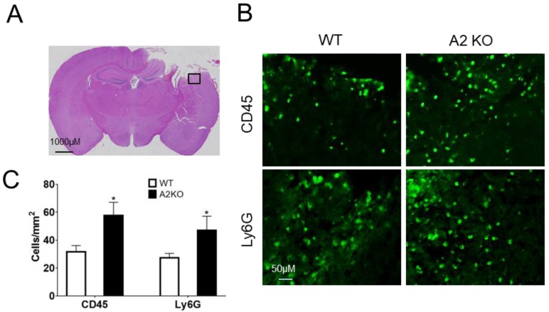
Figure 4.
AnxA2 deficiency enhances leukocytes brain infiltration after TBI. Brain tissues were collected at day two after TBI and sectioned for hematoxylin and eosin (H&E) staining and immunohistochemistry. (A) The black box in the representative H&E staining indicates the image field of CD45 and Ly6G-immunestaining in ipsilaternal cortex. (B) Representative CD45 and Ly6G-immunestaining images. (C) Quantitation of CD45 and Ly6G-positive cells in peri-lesion cortex of ipsilateral hemisphere. Data are expressed as mean ± SEM; n = 4, * p < 0.05 compared to WT.

