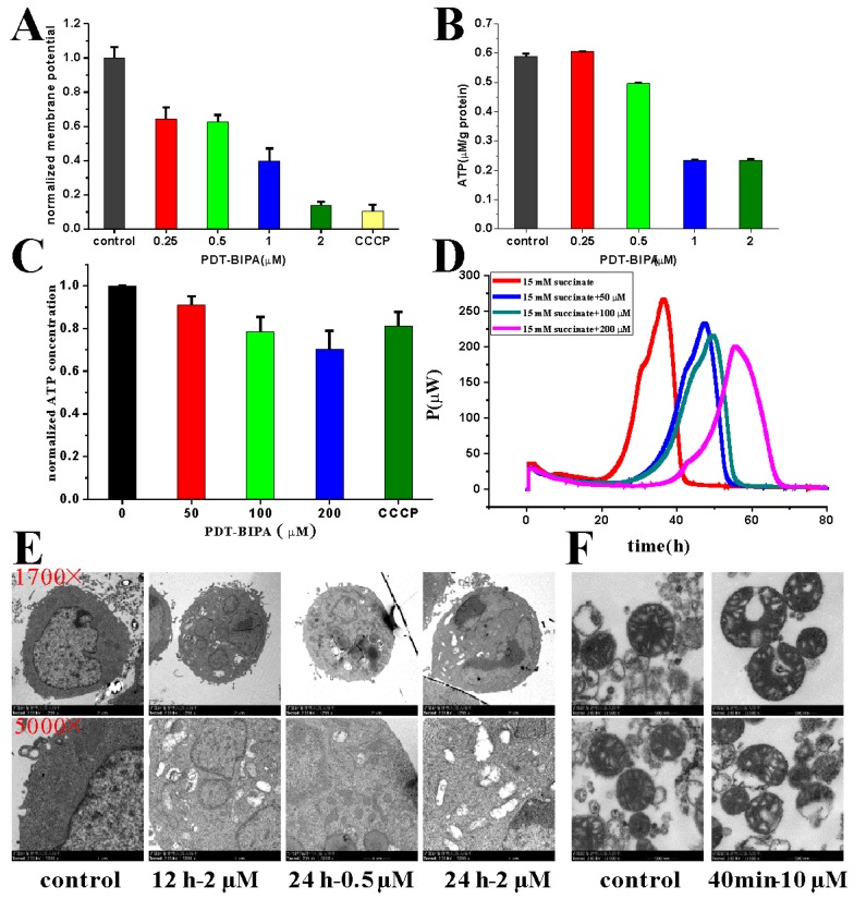
Figure 4.
The impairment of mitochondria caused by PDT-BIPA. The mitochondrial membrane potential of HL-60 cells was collapsed greatly where the CCCP group was the positive control (A). As a result, PDT-BIPA incubation caused the less ATP level of HL-60 cells (B) and the isolated mitochondria (C). As well, the heat output of isolated mitochondria was delayed (D). The cellular structure especially mitochondria was damaged as the incubation time and dosage increase (E). Besides, the isolated mice liver mitochondria demonstrated ruptured membrane under exposure of PDT-BIPA for 40 min (F). (the above and the below two pictures are the same group).

