Figure 1.
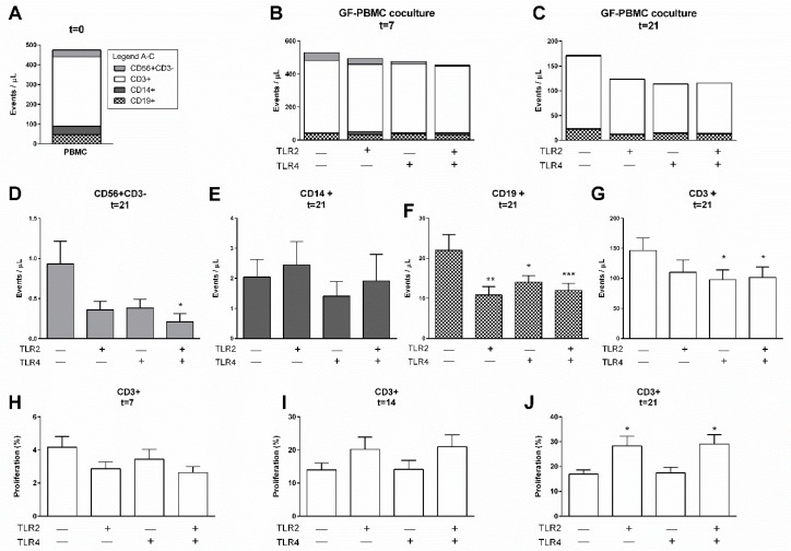
TLR agonists affect the survival of PBMCs in the presence of gingival fibroblasts. Characterization of heterogeneous cell populations: (A) immediately after isolation (t = 0), (B) after 7 days, and (C) after 21 days. No significant differences were found between total leukocyte cell numbers of control, TLR2, TLR4, and TLR2+TLR4 stimulated conditions for 7 nor for 21 days. Specification of cells from (C) at 21 days is shown for (D) CD56+CD3−, (E) CD14+, (F) CD19+, and (G) CD3+ cells, showing diminished numbers of CD56+, CD19+, and CD3+ cells and not CD14+ after TLR2 or TLR4 stimulation. The proliferation of CD3+ cells over time is presented as percentage divided cells at (H) 7, (I) 14, and (J) 21 days. Over time, a trend of increased proliferation was seen (note differences in values at y-axes). A significant increase in proliferation of CD3+ cells was seen in PBMC–GF cocultures with TLR2 agonists, after 21 days. Data are presented as events per microliter (A–G), + SEM (D–G), or in percentages + SEM (H–J). n = 6 GF donors. Statistical significance was calculated (D–J) using an ANOVA with multiple comparisons (Tukey post-hoc). Significant differences are shown in comparison to control conditions (without TLR agonists). * p < 0.05, ** p < 0.01, *** p < 0.001.

