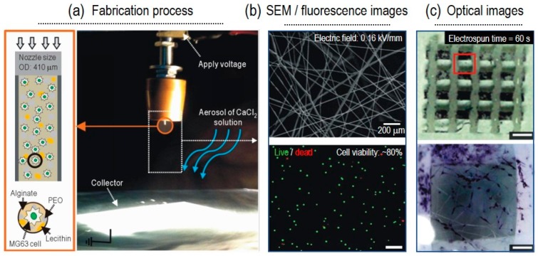
Figure 2.
(a) Schematic and optical image for fabrication process. (b) SEM/fluorescence (live/dead) images of fabricated cell (MG63)-laden electrospun fibers. (c) Optical images of alkaline phosphatase (ALP) stained cells. Figure adapted with permission from [21]. Copyright 2015 Elsevier.

