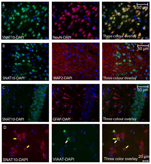
Figure 1.

(A) Mouse brain sections stained with anti-SNAT10 (green), anti-NeuN (red), and combined images (right most micrograph). Staining of cell bodies with SNAT10 is obvious, with an almost 100% overlap with the anti-NeuN antibody, although anti-SNAT10 staining appears more prominent close to the cell nuclei. (B) Mouse brain sections stained with anti-SNAT10 (green), anti-MAP2 (red) and combined images (right most micrograph). Staining of anti-SNAT10 and anti-MAP2 appears in the same cells, but with no in situ overlap, suggesting that SNAT10 is not expressed in any neuronal projections. (C) Mouse brain sections stained with anti-SNAT10 (green), anti-GFAP (red) and combined images (right most micrograph). No overlap between GFAP and SNAT10 is observed suggesting no expression of SNAT10 in GFAP positive glia cells. (D) Fluorescence micrograph of primary cell culture from adult mouse brain where the inhibitory neurons are marked with eGFP (green) and custom-made anti-SNAT10 antibody (Innovagen) with Alexa Fluor 594 (red). The nucleus is stained in blue using DAPI. Left most micrograph illustrates SNAT10 staining in red with two yellow arrows and the nuclei stained with DAPI are in blue. Middle micrograph with white arrow shows eGFP expressing inhibitory neuron in green and the nuclei in this micrograph are in blue. Right most micrograph shows a three color overlay with overlap between green and red staining indicating expression of SNAT10 in the inhibitory neurons with both yellow and white arrows. The yellow arrow indicates a cell marked with only red, revealing expression of SNAT10 in a neural cell other than inhibitory neuron.
