Figure 2.

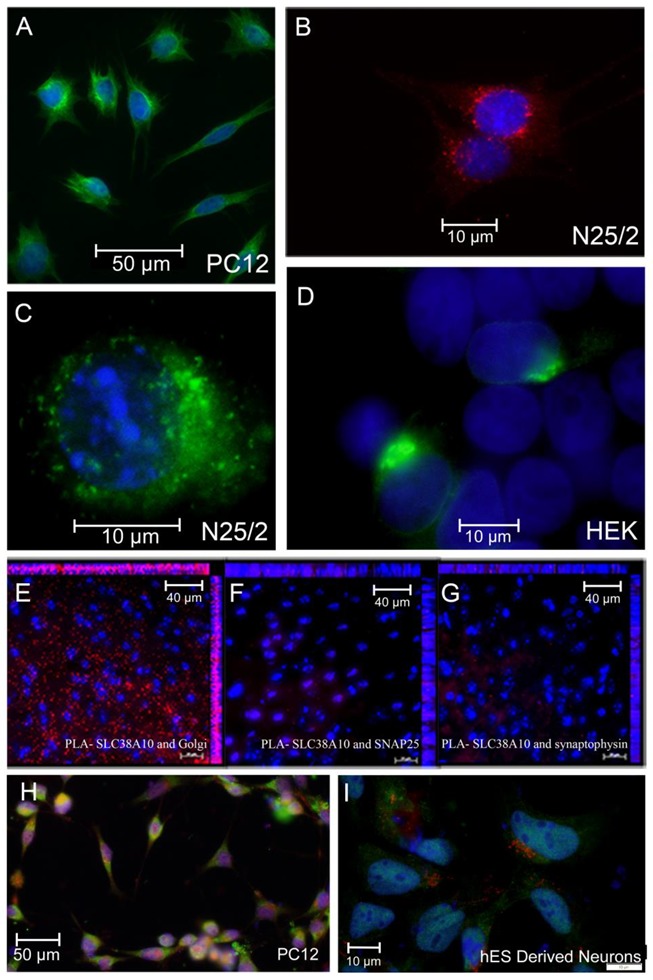
Fluorescence micrograph of PC12, N25/2, and HEK293 cells expressing SLC38A10 illustrating its intracellular localization adjacent to nuclei and proximity ligation assays revealing different degrees of interaction between SNAT10 and Golgi protein, SNAP25 and synaptophysin, respectively. (A) PC12 cells stained with custom-made SNAT10 antibody (Innovagen) with Alexa Fluor 488 (green) and nuclei stained with DAPI are shown in blue. (B) N25/2 cells stained with custom-made SNAT10 antibody (Innovagen) with Alexa Fluor 594 (red) and nuclei stained with DAPI are shown in blue. Using of two different secondary antibodies (Alexa Fluor 488 and Alexa Fluor 594) in (A,B) respectively nullifies any effect that the secondary antibodies might have had on the cell lines and therefore shows true localization of SNAT10. (C) Fluorescence micrograph of N25/2 cells transfected with SNAT10-eGFP construct showing localization of SNAT10 adjacent to the nuclei that are stained with DAPI and shown in blue. (D) Fluorescence micrograph of HEK293 cells transfected with SNAT10-eGFP construct showing localization of SNAT10, adjacent to the nuclei that are stained with DAPI and shown in blue. Both (C,D) show the expression of SNAT10 at one of the edges of the nuclei. (E) Micrograph visualizing proximity ligation assay (PLA) signals in red between SNAT10 and Golgi protein. (F) Micrograph visualizing PLA signals in red between SNAT10 and SNAP25. (G) Micrograph visualizing PLA signals in red between SNAT10 and synaptophysin. In (E–G) nuclei are stained with DAPI and shown in blue. The micrographs (E–G) show a comparison between Golgi protein, SNAP25, and synaptophysin in relation to SNAT10. It is obvious that SNAT10 is in proximity to Golgi proteins and not SNAP25 or synaptophysin. (H) PC12 cells transfected with a FLAG-tagged SNAT10 construct and stained with anti-FLAG antibody (green) and Golgi protein (red) as a marker for the Golgi apparatus. (I) Human embryonic stem cell-derived neurons transfected with a FLAG-tagged SNAT10 transgene and stained with anti-FLAG antibody (green) and Golgi protein (red).
