Figure 1.
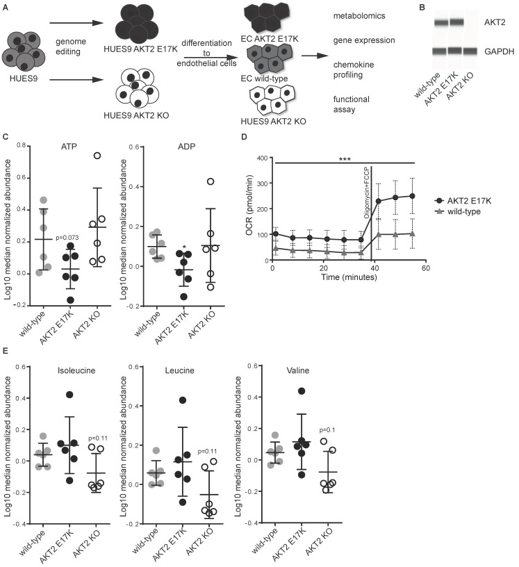
Metabolic dysregulation of human pluripotent stem cell (hPSC) endothelial cells (ECs) carrying AKT2 mutations. (A) Schematic representation of the engineered endothelial cells and a list of the subsequent experimentation. (B) Western blot of AKT2 and GAPDH from hPSC-EC cell lysates. (C) Abundance of ATP and ADP from six replicates of each hPSC-EC as measured by mass spectrometry from cellular lysates ± SD. (D) Cellular oxygen consumption rate under basal conditions and after stimulation with oligomycin (2 μM) and carbonyl cyanide-p-trifluoromethoxyphenylhydrazone (FCCP) (0.5 μM) of AKT2 wild type (WT) and AKT2 E17K hPSC-ECs. Data are presented as mean ± SD of 10 measurements per group per timepoint. (E) Abundance of branched-chain amino acids (BCAAs) from six replicates of each hPSC-EC as measured by mass spectrometry ± SD. For all experiments in this figure, * p < 0.05, *** p < 0.001.

