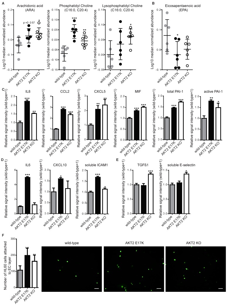
Figure 2.
Inflammation is induced in hPSC-ECs carrying either of the two AKT2 mutations. For each experiment, hPSC-ECs with AKT2 mutations (AKT2 E17K and AKT2 knockout (KO)) were compared to WT. (A) Abundance of proinflammatory lipids from six replicates of each hPSC-EC and (B) an anti-inflammatory lipid, eicosapentaenoic acid, as measured by mass spectrometry in cellular lysates ± SD. (C–E) Inflammatory mediators measured using a multiplexed sandwich immunoassay and compared to WT with (C) showing those induced in both AKT2 E17K and AKT2 KO, (D) those induced only in AKT2 E17K, and (E) those induced only in AKT2 KO. (F) Quantification of leukocyte-like cells (HL-60) adhering to hPSC-ECs presented as the mean of triplicate experiments ± SD. * p < 0.05, ** p < 0.01, *** p < 0.001. Representative images for each cell line are shown.

