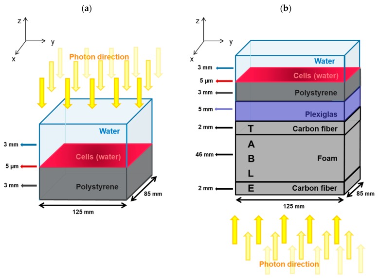
Figure 6.
(a) Setup used for simulation for SARRP irradiation. The cell culture chamber was modeled by three layers of material (from top to bottom): 3 mm of liquid water representing the cell culture medium, 5 µm of liquid water defining the cell layer, and 3 mm of polystyrene modeling the chamber material. The dimensions in the x–y plane of each layer were 125 mm × 85 mm. Photons were generated in a parallel beam emitted from above and directed towards the cell monolayer. (b) Setup for simulation of medical linear accelerator irradiation. The cell chamber (same as the one used in the SARRP simulation) was placed on a table made of carbon fiber and foam (the carbon fiber was 4 mm high and the foam was 46 mm high). Between the cell chamber and the table, a layer of 5 mm in height of Plexiglas® was placed to ensure an electronic equilibrium. Photons were generated in a parallel beam from below the table towards the cell chamber.

