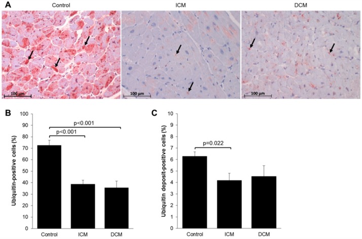
Figure 2.
(A) Immunohistochemical staining for ubiquitin on sectioned myocardium from control (n = 10), ICM (n = 11), and DCM (n = 10) patients. Arrows denote ubiquitin deposits. Quantification of ubiquitin staining of (B) ubiquitin-positive cells and (C) ubiquitin deposit-positive cells. Results are expressed as mean ± SEM.

