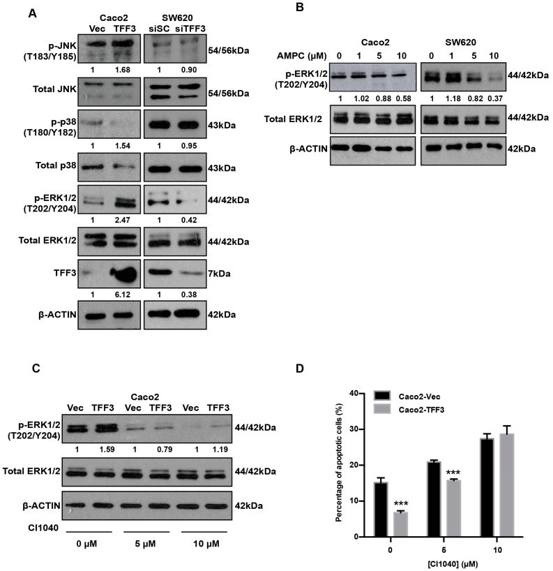
Figure 6.
TFF3 activates the ERK1/2 (p44/42 MAPK) pathway in CMS4 CRC cells. (A) Western blot analysis of phosphorylated and total MAPKs in Caco2-Vec and Caco2-TFF3 cells and SW620-siSC and SW620-siTFF3 cells. β-ACTIN was used as input control. (B) Caco2 and SW620 cells were treated with the indicated concentrations of AMPC (with DMSO as vehicle) for 24 h. Levels of p-ERK1/2 and expression of total ERK1/2 were detected by western blot analysis. β-ACTIN was used as an input control. (C) Caco2-Vec and Caco2-TFF3 cells were treated with the indicated concentrations of MEK inhibitor (CI1040) or DMSO (vehicle control) for 24 h. Levels of p-ERK1/2 and expression of ERK1/2 were detected by western blot analysis. β-ACTIN was used as an input control. (D) Annexin V/PI staining analysis was performed to determine apoptosis in Caco2 stable cells. ***, p < 0.001.

