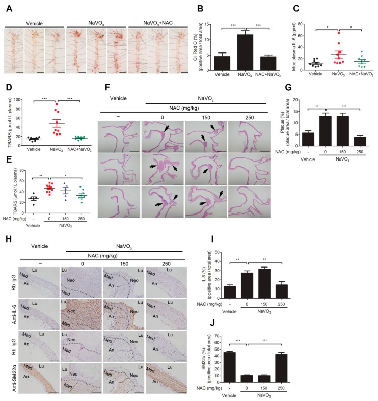
Figure 6.
Anti-oxidant N-acetylcysteine prevents NaVO3-induced atherosclerosis in ApoE−/− mice. ApoE−/− mice administered NaVO3 (4 mg/kg) once a week were injected intraperitoneally with saline (vehicle) or NAC (250 mg/kg) three times weekly for 12 weeks (n = 9 vehicle; n = 9 NaVO3; n = 8 NaVO3 + NAC). (A) Lipid contents in aorta were analyzed by Oil Red O staining. Scale bars represent 5 mm. (B) The area of positive staining for Oil Red O was quantified using ImageJ software as a percentage of total aortic area. (C) Plasma IL-6 levels were measured by ELISA. (D) Plasma ROS levels were measured by TBARS assay. (E–J) ApoE−/− mice administered NaVO3 (4 mg/kg) once a week were injected intraperitoneally with different amounts of NAC (150 or 250 mg/kg) three times weekly for 12 weeks (n = 5 vehicle; n = 10 NaVO3; n = 5 NaVO3 + 150 mg/kg NAC; n = 8 NaVO3 + 250 mg/kg NAC). (E) Plasma ROS levels were measured by TBARS assay. (F) Paraffin-embedded tissues with atherosclerotic plaque (black arrow) were observed by hematoxylin and eosin stain at 40× magnification. Scale bars represent 1 mm. (G) Atherosclerotic plaque in paraffin-embedded aorta tissues were quantified using ImageJ software as a percentage of total aortic area in each section. (H) IL-6 and SM22α contents in aorta tissues were analyzed by immunohistochemistry. Immunopositive areas are shown at 400× magnification. An: adventitia; Lu: lumen; Neo: neointima; Med: media. Negative control represents staining with an isotype control antibody. Scale bars represent 100 µm. (I) IL-6 and (J) SM22α immunopositive areas in paraffin-embedded aorta tissues were quantified using ImageJ software as a percentage of total aortic area in each section. Data represent mean ± SEM. The solid black line denotes the mean value. * p < 0.05; ** p < 0.01; *** p < 0.001.

