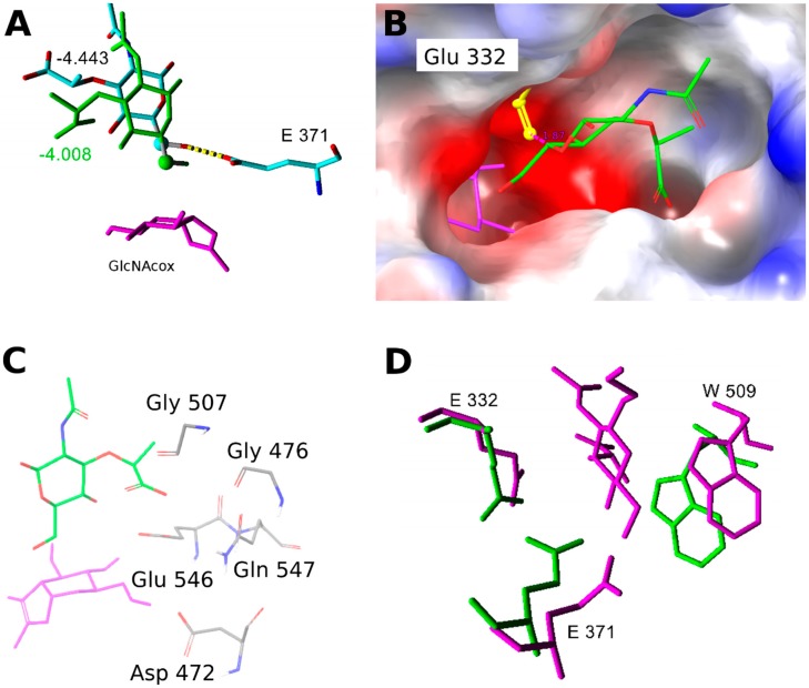
Figure 7.
(A) Orientation of MurNAc in the WT-GlcNAcox complex: green—close to the transglycosylation donor; element color—far, corresponds to site 2. The respective Glide absolute binding scores are shown in the figure. GlcNAcox is shown in magenta, hydrogens are hidden, and the C-6 atom is represented by a ball. (B) Electrostatic potential surface of the WT enzyme active site with GlcNAcox (magenta) and the MurNAc acceptor (element color). Orientation of Glu332 (yellow) in one of the analyzed snapshots disallows a close interaction between the acceptor and donor. The negatively charged environment is shown in red, and that which is positively charged is presented in blue. (C) Residues with a negative charge in the vicinity of the MurNAc acceptor carboxyl group when it binds close to the transglycosylation donor. (D) Overlay of the aglycone binding site residues of WT-GlcNAcox (green) and Y470H-GalNAcox (magenta) with docked MurNAc-OPr. Residues with similar side chain orientations are hidden.

