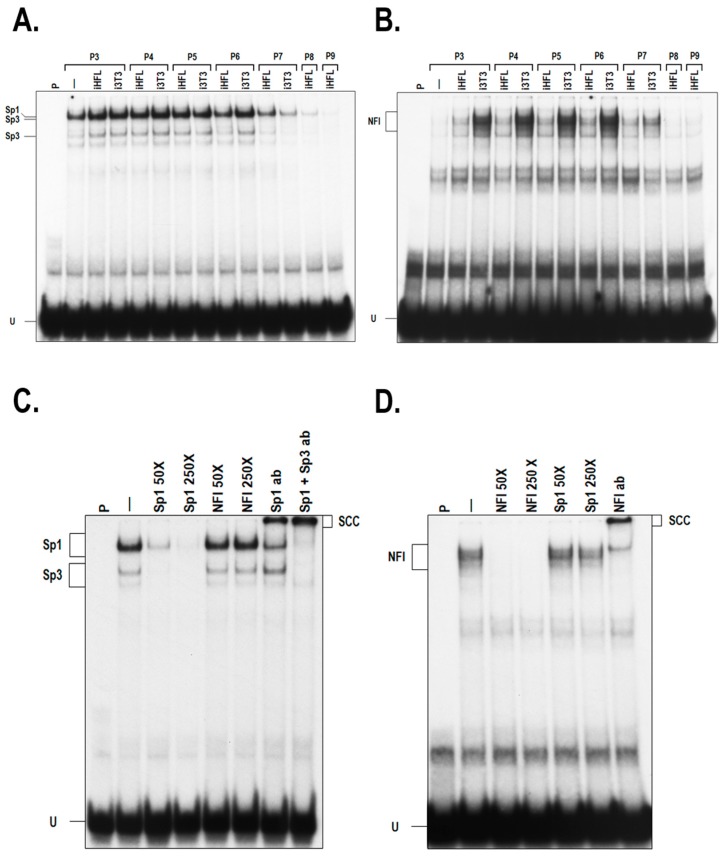
Figure 3.
DNA binding properties of Sp1 and NFI in hCECs cultured with a feeder layer. (A) EMSA (Electrophoretic Mobility Shift Assay) showing the DNA binding capacity of Sp1 in hCECs (hCEC52 cell population) cultured with i3T3, iHFL, or without a feeder layer (-) over culture passages. (B) Nuclear extracts from panel A were used to evaluate the capacity of NFI to bind to its high affinity target site in EMSA. (C,D) Nuclear proteins from hCECs grown with iHFL at passage 3 were incubated with either the Sp1 (panel C) or NFI (panel D) labeled probe in the presence of a 50- or 250-fold molar excess of an unlabeled oligonucleotide bearing either the Sp1 or the NFI high affinity-binding site as competitor. Formation of DNA-protein complexes was then monitored by EMSA. When indicated, antibodies directed against Sp1 (Sp1 ab), Sp3 (Sp3 ab) or NFI (NFI ab) were added to the reaction mix prior to the EMSA. The position of the Sp1 and NFI complexes is indicated, as well as that of their corresponding supershifted complexes. P: labeled probe alone; -: labeled probe incubated with nuclear proteins but without unlabeled competitor; U: unbound fraction of the labeled probe.

