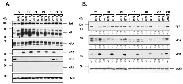
Figure 4.
Western blot analysis of Sp1 and NFI in hCECs cultured with a feeder layer. (A) Nuclear extracts prepared from hCECs (hCEC52 cell population) grown in the presence of either i3T3 or iHFL at different passages (P3 to P9) were separated by SDS-PAGE and Western blotted using antibodies raised against Sp1, NFI-total (NFI), NFIA, NFIB, NFIC and NFIX. (B) Western blot analysis of Sp1, NFIA and NFIB in hCECs grown alone (-) or with an i3T3 or iHFL feeder layer, in the presence of either no (0H) or 50µL/mL cyclohexemide for various periods of time (1 to 24 h). As a control, cells were also incubated for 24 h with the vehicle (DMSO). The position of the appropriate molecular mass markers is indicated (kDa). Values shown beneath each blot correspond to the ratio of the TF signal over that of actin.

