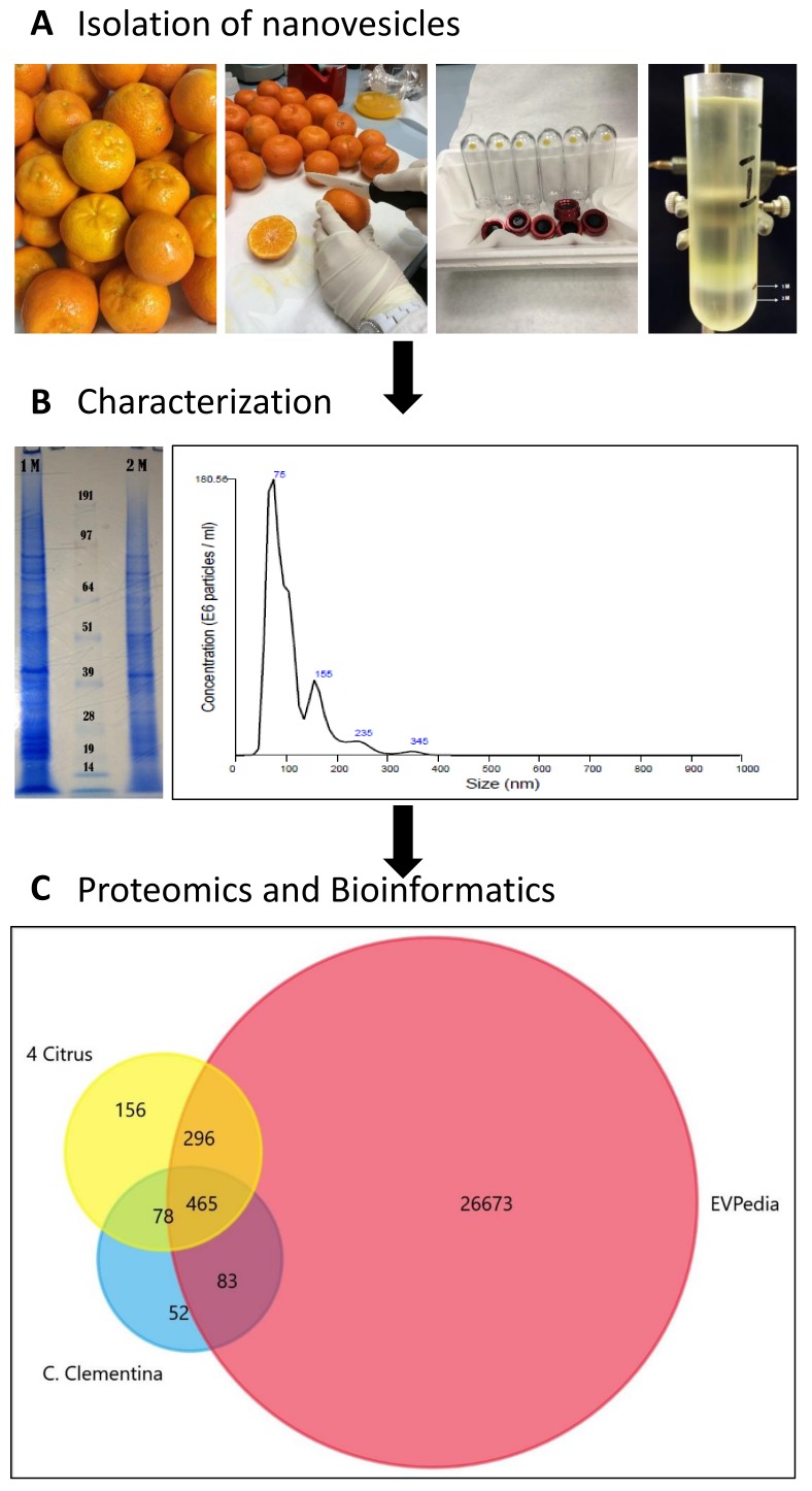
Figure 1.
Schematic chart of the experimental work performed to isolate, characterize and analyze C. clementine fruit juice-derived exosome-like nanovesicles. (A) Lower left image shows the pellets obtained after diffferential ultracentrifugation (UC) lower right image shows the separation obtained by sucrose/D2O double cushion UC. The vesicles floating above the 1M sucrose/D2O cushion were found to be similar in density to mammalian extracellular vesicles. (B) sodium dodecyl sulfate polyacrylamide gel electrophoresis (SDS-PAGE) protein profiles (right) and of vesicle populations in the 1 M and 2 M sucrose/D2O cushions and the particle-size distributions of vesicles isolated in the 1M sucrose/D2O cushion and measured using nanoparticle tracking analysis (NTA). (C). Venn diagram generated by FunRich software [1] shows the numbers of unique and common Orthologous Groups (OGs) of the identified protein. OGs of Citrus clementina (azure) were compared to four citrus species (C. sinensis, C. limon, C. paradise and C. aurantium) (yellow) [13] and EVpedia (red) [14].

