Figure 3.
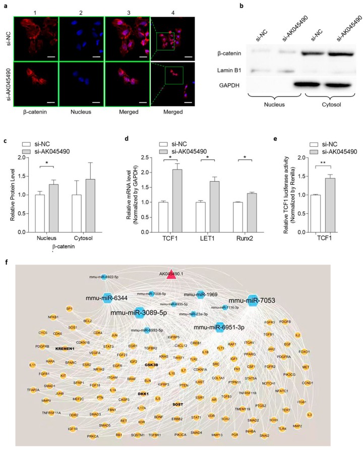
Knockdown of AK045490 promoted β-catenin nuclear translocation and up-regulated the expression of TCF1, LEF1, and Runx2. (a) IHF staining showed the location of β-catenin in the cells transfected with AK045490-siRNA (si-AK045490) or scrambled-control-siRNA (si-NC), respectively. β-catenin was stained as red and nuclei were stained by DAPI showing blue. Bar (1, 2, 3): 20 µm. Bar (4): 50 µm. (b) Representative western blots of the nuclear translocation of β-catenin. The nuclear (nucleus) and cytosolic (cytosol) fractions of proteins isolated from AK045490 knockdown cells and control cells were probed for β-catenin. Lamin B1 and GAPDH were used as internal controls for nuclear and cytosol fractions, respectively. Full unedited gels available in the Supplementary file. (c) Quantification of nuclear and cytosol levels of β-catenin with Lamin B1 and GAPDH as internal control, respectively. (d) mRNA expression of TCF1, LEF1, and Runx2 as detected by real time PCR. (e) TCF1 activity in MC3T3-E1 cells transfected with AK045490 siRNA, as detected by TOPflash luciferase reporter assay. (f) Pattern diagram showed the network of lncRNA–microRNA–RNA interaction. All data were expressed as mean ± SD. Student’s t-test was performed for comparison between two groups. p values less than 0.05 were considered significant in all cases (* p < 0.05, ** p < 0.01). n = 3 in each group.

