Figure 5.
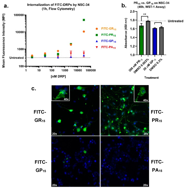
Arginine-rich fluorescein isothiocyanate-labelled dipeptide repeat proteins (FITC-DRPs) enter cells in a fast, quantifiable dose-dependent manner. Mean fluorescence intensity (MFI) values for FITC-DRP-treated mouse spinal cord x neuroblastoma hybrid cells (NSC-34) were generated from a gated region (Supplementary Figure S6) selecting only for values greater than untreated cell fluorescence values (dotted line). Data indicates detectable FITC-poly-glycine-arginine (GR15) (orange) and FITC-poly-proline-arginine (PR15) (green) uptake by NSC-34 at doses as low as 3 nM. Minimal entry of FITC-poly-glycine-proline (GP15) (blue) by 30 μM, and little to no entry of FITC-poly-proline-alanine (PA15) (red) was observed. Data are presented as mean values ± standard error of the mean (error bars) (a). Using dose-pairing of PR15 and GP15 based on flow cytometry data, a WST-1 assay indicated significant toxicity of PR15 (red) and not GP15 (blue) compared to Dimethyl Sulfoxide (DMSO) controls (gray). Data are presented as mean values ± standard deviations (error bars) ** denotes p < 0.01 (comparing DRP-dosed to corresponding DMSO control by one-way ANOVA analysis of variance followed by Dunnett’s test) (b). Arginine-rich FITC-DRPs localize differently in NSC-34. Confocal laser scanning microscopy of FITC-DRP treated NSC-34 reveals distinct localization of arginine-rich DRPs and limited entry of non-arginine-rich DRPs. Nuclei are 4’6-diamidino-2-phenylindole (DAPI)-stained, and FITC-DRPs appear in green. Cells were imaged using 20× and 40× objectives as indicated by white text (c). Additional 3-D images, as well as flow cytometry after 24 h incubation, are available as supplementary material (Supplementary Figures S1, S2, S4 and S5).

