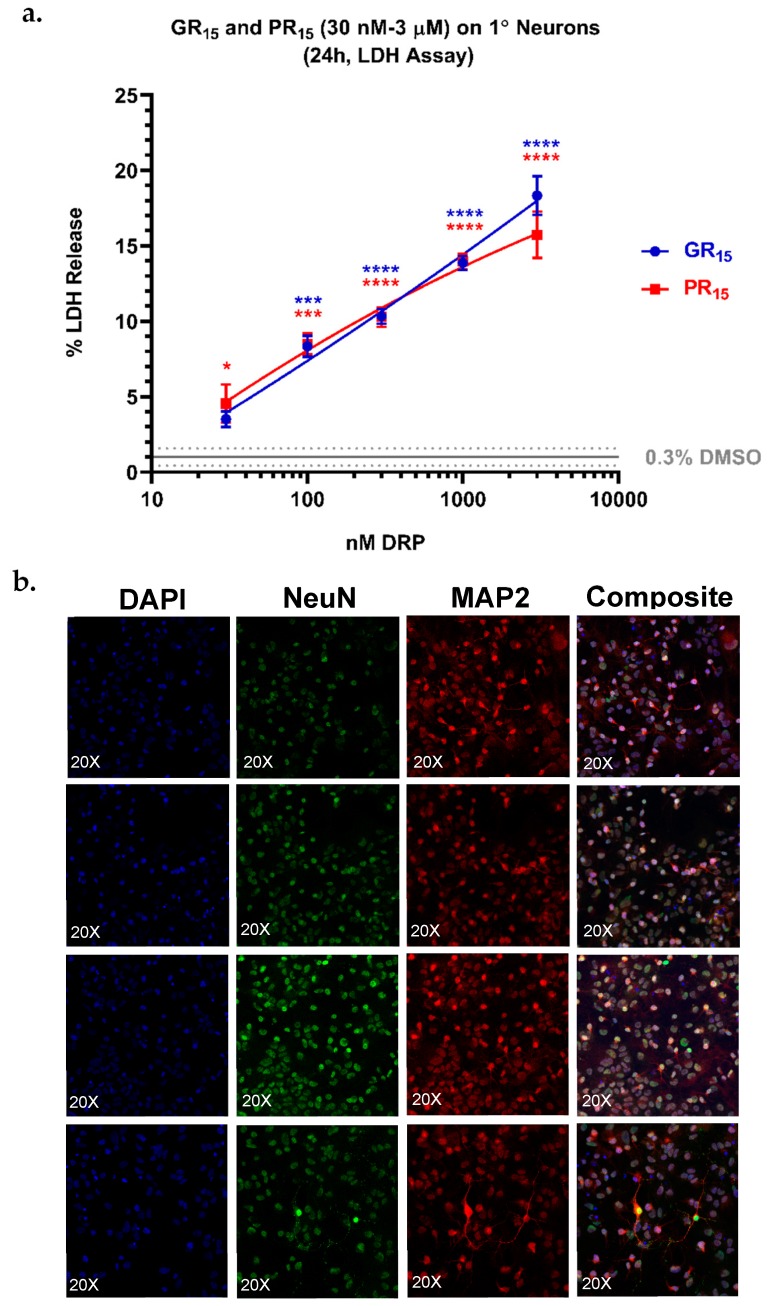
Figure 7.
Lactate dehydrogenase (LDH) assay on primary (1°) neurons revealed an increased sensitivity to arginine-rich dipeptide repeat proteins (DRPs). 1° neurons treated with poly-glycine-arginine (GR15) or poly-proline-arginine (PR15) exhibited significant, dose-dependent LDH release, with a maximal signal of 20% LDH release at 3 µM DRP doses (a). Co-staining of nuclei, neuronal nuclei, and neuron cytoskeletons with 4’6-diamidino-2-phenylindole (DAPI) and antibodies against hexaribonucleotide binding protein-3 (NeuN), and microtubule-associated protein 2 (MAP2) revealed a high purity of the 1° neuron population tested (b). Data are presented as average % values ± standard deviations (error bars). % values indicate the % LDH release DRP-treated cells exhibited where 0% LDH release reflects a negative control of untreated 1° neurons, and 100% LDH release reflects a positive control of lysed 1° neurons. Average % LDH release induced by the highest concentration of solvent tested (0.3% DMSO) is indicated with a gray line, with gray dotted lines representing the standard deviation of this value. * denotes p < 0.05, *** denotes p < 0.001, and **** denotes p < 0.0001 (comparing DRP-treated values to DMSO-treated values at each dose by one-way ANOVA analysis of variance followed by Dunnett’s test). Data are presented as mean values ± standard deviations (error bars).

