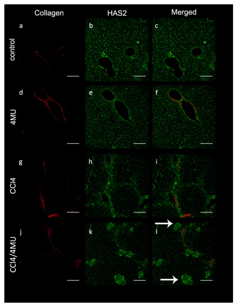
Figure 2.
HAS2-positive cells accumulation around collagen fibers in CCL4-exposed animals and aggregate in clamps in the CCL4/4MU-treated group (see arrows). The second harmonic generation method was used for collagen detection. (a,d,g,j). HAS2 was detected immunohistochemically (b,e,h,k). Merged images (c,f,i,l), scale bar 100 um. Arrows indicates clamps of HAS2-positive cells.

