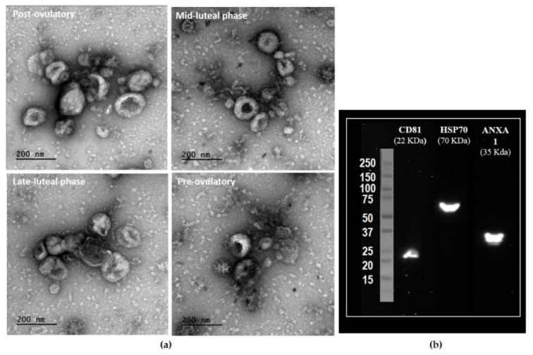
Figure 1.
Characterization of bovine oviductal extracellular vesicles (oEVs). Representative images of exosomes (30–100 nm) and microvesicles (>100 nm) in oEV preparations observed by transmission electron microscopy (TEM) across the estrus cycle (a) and Western blotting characterization of bovine oEVs for known exosomal protein markers (b). A pool of samples from four different stages was used, showing that oEVs were positive for CD81, HSP70, and ANXA1.

