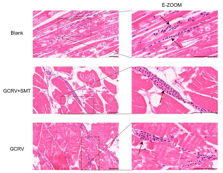
Figure 6.
The vascular injury was inhibited when inhibiting the function of iNOS by SMT. H&E-stained sections of the vessel in different treatments (scale 50 μm). The vascular wall was broken and the infiltration of blood cells could be observed in the GCRV group. The vascular wall was complete in GCRV+SMT and blank. The black arrow shows the vessel and the white shows hemorrhage.

