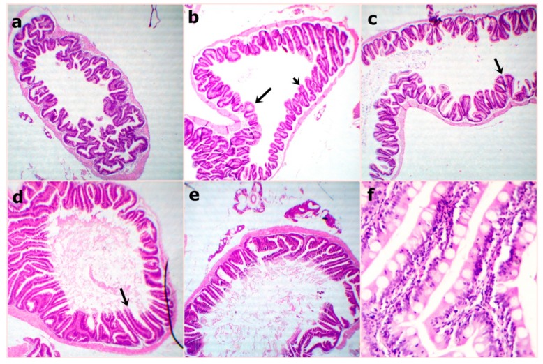
Figure 2.
Representative photomicrograph of H&E stained sections from fish intestine at the end of the experiment showing (a) WPC0–reduced villus height and width at 40x; (b) WPC13.8—distinctly arranged tall villi at 40x; (c) WPC27.7—marked tall and branched villi with some broad tips (arrow) at 40x; (d) WPC41.6—marked very tall and thin villi (arrow) at 40x; (e,f) WPC55.5—clearly tall and thin villi with partially damaged tips (arrow) with reduced goblet cell count and inter-epithelium lymphocytic infiltration (arrow) at low 40x and high magnification 400x.

