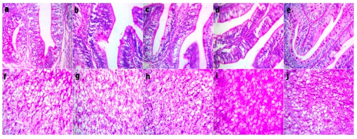
Figure 6.
Representative photomicrograph of the Periodic Acid Schiff stained sections at magnification 400x from the fish intestine and liver before bacterial challenge showing the red stainable intestinal goblet cells and mucus within the limit and no difference between all groups. Red granular stainable materials (glycogen) distributed in the hepatocyte cytoplasms are within the normal limit, but they are mildly increased in the WPC41.6 and WPC55.5 liver sections. (a,f) WPC0, (b,g) WPC13.8, (c,h) WPC27.7, (d,i) WPC41.6, (e,j) WPC55.5.

