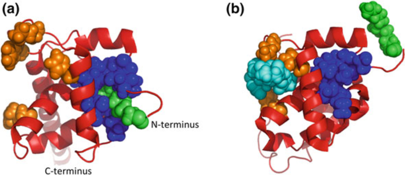
Fig. 5.
The MA myristyl switch. HIV-1 MA exists in two conformations: a In the cytoplasm, the hydrophobic myristic acid moiety is sequestered into a groove on the surface of the protein. b At the plasma membrane, MA binds to PI(4,5)P2 (di-C4-PI[4,5]P2 in this structure), causing a conformational shift and exposure of myristic acid. a and b were generated using Pymol and PDB coordinates 2H3I and 2H3Q, respectively (Saad et al. 2006). MA, red; myristic acid, green; myristate-binding groove, blue; PI(4,5)P2 binding residues, orange; di-C4-PI(4,5)P2, cyan

