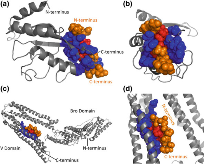
Fig. 6.
Late-domain peptide binding to ESCRT proteins. a TSG101 ubiquitin E2 variant (UEV) domain bound to PTAP peptide. b View of UEV–PTAP interaction, facing into the binding groove. c ALIX V domain bound to YPLTSL peptide. d Close-up view of the ALIX-YPLTSL binding site. Host proteins are shown in gray, with binding sites in blue. Late-domain peptides are shown in orange, with interacting residues in red. Structures of late-domain interactions with TSG101 and ALIX are generated using Pymol with PDB coordinates 1M4P and 2RO2, respectively (Pornillos et al. 2002; Zhai et al. 2008)

