Fig. 2.
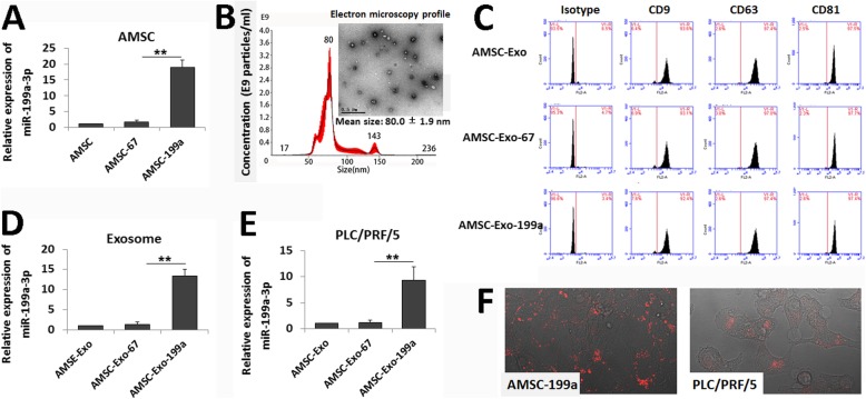
AMSC-Exo-mediated miR-199a-3p transfer into HCC cells. a Real-time PCR detection of miR-199a-3p expression in navie AMSCs and miRNA-modified AMSCs. b Transmission electron microscopy detection on AMSC-Exo morphology and nanoparticle tracking analysis on exosome particle size and concentration (Scale bar = 0.5 μm). c Flow cytometric analysis of the surface markers of the exosomes derived from navie AMSCs and miRNA-modified AMSCs. d Real-time PCR detection of miR-199a-3p expression in AMSC-Exo. e Real time-PCR detection of miR-199a-3p expression in AMSC-Exo-treated PLC/PRF/5 cells. f Confocal images of AMSC-199a stained with DilC16(3) and the PLC/PRF/5 cells incubated by the exosomes derived from DilC16(3)-stained AMSC-199a. Data are presented as mean ± S.D. (**P < 0.01, n = 3)

