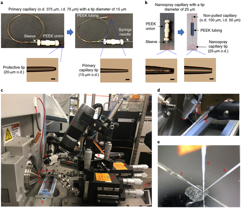
Fig. 2 |. Platform for high-resolution imaging of biological tissues using nano-DESI MSI.
a, Fabrication of the primary capillary. Top panel shows the key stages for fabricating the primary capillary. Bottom panels show photos of the pulled tips. Scale bars, 25 μm. b, Fabrication of the nanospray capillary. Top panel shows the key stages for fabricating the nanospray capillary. Bottom panels show photos of the pulled tips. Scale bars, 25 μm. c, Photo of the imaging platform, highlighting (1), instrument mounting flange; (2), xyz 500 inline micro-positioner; (3), primary capillary; (4), nanospray capillary; (5), shear force probe; (6), sample holder; (7), Dino-Lite microscope; and (8), motorized micro-positioner (for shear force probe). d, Zoomed-in picture of the probes, corresponding to the red dashed box in c. e, A photo of the probes and sample recorded by a side-view microscope during the imaging experiment.

