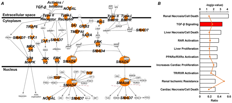
Figure 8: Pathway analysis of miR-146b-5p targets suggests important role for TGF-β signaling.
Predicted targets of miR-146b-5p were analyzed using IPA software to identify over-represented canonical pathways and provide strong rationale for further investigation. Both the (A) canonical TGF-β signaling pathway and the (B) TGF-β toxicity list (i.e. biological functions associated with pathological outcomes) had a disproportionate representation of miR-146b-5p targets. (A) Each node within the canonical TGF-β pathway containing a predicted target is highlighted orange. (B) The top axis (bar graph) represents p-value associated with the toxicity list based on target representation and the bottom axis (orange line) represents the ratio of molecules within the given toxicity list that are miR-146b-5p targets.

