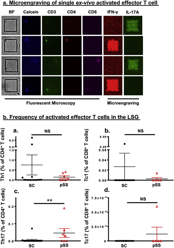
Fig. 1.
Microengraving shows greater infiltration by activated Th17 cell in the labial salivary glands of pSS patients. a) Microengraving of single ex-vivo activated effector T cell. Representative fluorescent microscopy coupled with microengraving of secreted cytokines from isolated individual T cell. Fluorescent antibody staining was performed with anti-CD3-FITC (green) anti-CD4-PE (red), anti-CD8-APC (Magenta), and Calcein violet-405 (blue), a marker of viable cells. Secreted cytokines were captured during microengraving and detected with anti-IFN-γ (red) and anti-IL-17A (green). b) Quantification of activated effector T cells isolated from the LSG of SC subjects () and pSS patients () expressing (a) CD3+CD4+IFN-γ+(Th1), (b) CD3+CD8+IFN-γ+(Tc1), (c) CD3+CD4+IL-17+ (Th17), and (d) CD3+CD8+IL-17+ (Tc17). The frequency in percentage was determined by using the percentage (multiplied by 100) of the total number of Th1, Th17, Tc1, and Tc17 cells from wells with single live cells among the total number of wells with single CD4+ or CD8+ cells. Statistics were performed using an unpaired two-tailed Mann-Whitney test. Significance was determined as **p < 0.01, and NS: not significant.

