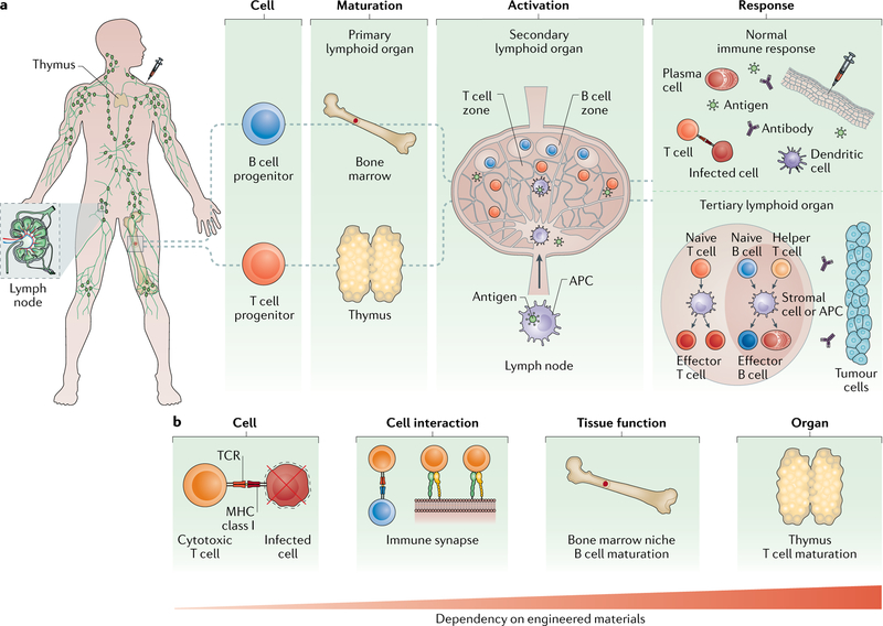
Fig. 1 |. The different levels of the immune response.
a | B and T cells originate in lymphoid organs and reside in the lymphatic system. During an immune response, Band T cells are first generated in primary lymphoid organs — the bone marrow and thymus — from haematopoietic stem cells (HSCs) and lymphoid progenitors. B and T cells then migrate to secondary lymphoid organs, such as the lymph node, where they localize in specific T cell and B cell zones. In these zones, each cell is activated by intact antigens or processed antigens presented on antigen-presenting cells (APCs), followed by differentiation into effector cells. B effector cells, such as plasma cells, and T effector cells, such as cytotoxic T cells, then migrate to sites of infection, including sites created by vaccine delivery. APCs, such as dendritic cells, encounter antigen at the infection site and present it to naive B and T cells in the lymph node for stronger and sustained responses. During disease, the normal immune response can be deregulated, leading to the formation of ectopic tertiary immune organs, which are structured immune aggregates often found near tumours. b | The immune response is regulated at the cell, tissue and organ levels. Efficient immune cell effector function is crucial for the targeting and killing of infected cells or tumour cells. Interactions between T cells and APCs (that is, the formation of immune synapses) play key roles in immune cell activation. Maturation of immune cells occurs in bone marrow niches and in the thymus. Engineering approaches are needed to recapture functionality at each biological scale. The dependency on material incorporation increases with the complexity of the immune function. MHC, major histocompatibility complex; TCR, T cell receptor.

