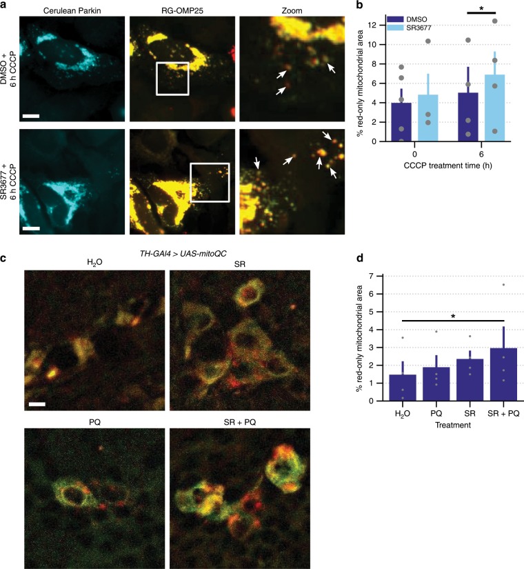
Fig. 3. Targeting of mitochondria to lysosomes is increased by SR3677.
a HeLa cells were co-transfected with Cerulean-Parkin and RG-OMP25. After 24 h, cells were pre-treated with either 0.5 µM SR3677 or DMSO for 2 h prior to induction of mitophagy with 10 µM CCCP treatment, in combination with E-64 and leupeptin. Red-only signal represents mitochondria localized to lysosomes, where GFP signal is quenched. Scale bars, 10 µm. b Quantification of the percentage of red-only mitochondrial area divided by the total non-background area (n = 4 independent experiments). P-values were determined by one-tailed paired Student’s t-test. c Seven-day-old TH-GAL4>UAS-mitoQC male flies were placed into vials containing the indicated treatments. Representative images of the dopaminergic neurons of TH-GAL4>UAS-mitoQC flies following feeding on fly food supplemented with H2O, 0.5 mM SR3677 (SR) or H2O/SR3677 combined with 5 mM paraquat (PQ). Scale bars, 10 µm. d Quantification of the percentage of red-only mitochondrial area divided by the total non-background area, averaged across 0.8-µm z-stacks. Data are expressed as mean ± s.e.m (n = 4 independent experiments). P-values were determined by one-tailed paired Student’s t-test, *P < 0.05. Error bars represent s.e.m.

