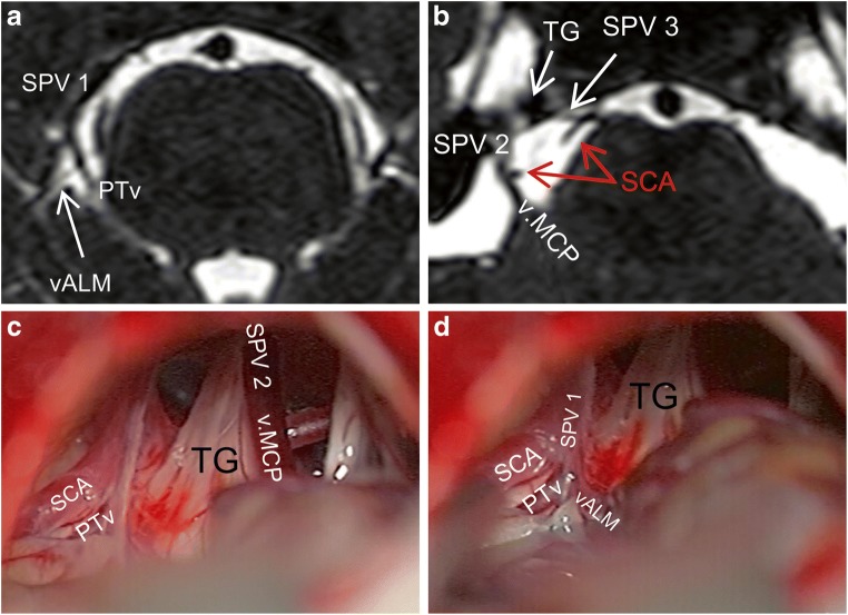
Fig. 7.
A case of right-sided CPA. a Axial bFFE magnetic resonance imaging showing an SPV1 composed of an ALMv and a PTv. b Another axial section showing an SPV 2 as the continuation of the v.MCP as well as an SPV 3 as a continuation of an TPv. c, d Corresponding intraoperative photographs of the same case. We can see here all the above described SPVC components, except the SPV 3 and its single tributary (TPv). This is because of the very medial course of the TPv and medial type III drainage of the SPV 3 into the SPS beyond the microsurgical field. ALMv anterior lateral marginal vein, bFFE balanced fast field echo, CPA cerebellopontine angle, SCA superior cerebellar artery, SPV superior petrosal vein, SPVC superior petrosal vein complex, SPV1 first SPV, SPV2 second SPV, SPV3 third SPV, TG trigeminal nerve, TPv transverse pontine vein, v.CPF vein of cerebellopontine fissure, v.MCP vein of the middle cerebellar peduncle

