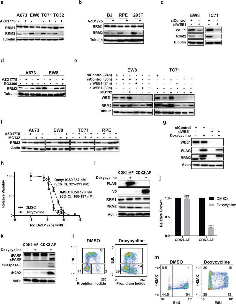
Figure 5.
CDK2 activation in Ewing sarcoma cells promotes RRM2 degradation. (A) Four Ewing sarcoma cell lines were treated with AZD1775 (500 nM) for 8 h. Cellular lysates were then harvested for immunoblotting for RRM1 and RRM2. (B) Three non-Ewing sarcoma cell lines were treated with AZD1775 as described in (A). (C) EW8 and TC71 cells were transfected with siRNA targeting WEE1. Cellular lysates were then collected 24 h after transfection for immunoblotting for WEE1 and RRM2. (D) EW8 and TC71 cells were treated with AZD1775 (500 nM), RO-3306 (10 μM), or the combination of AZD1775 and RO-3306 for 6 h. Cellular lysates were then harvested for immunoblotting for RRM2. (E) EW8 and TC71 cells were transfected with siWEE1 or siControl. MG132 (1 μM) was added 24 h after transfection. Cellular lysates were collected at 24 and 30 h after transfection for immunoblotting for RRM2 and WEE1. (F) A673, EW8, TC71, and RPE-tert cells were treated with AZD1775 (500 nM), MG132 (1 μM), or the combination of AZD1775 (500 nM) and MG132 (1 μM) for 6 h. Lysates were then collected for immunoblotting. (G) TO-FLAG-RRM2-T33A cells were treated with doxycycline in combination with siControl or siWEE1 for 24 h. Cellular lysates were then collected for immunoblotting. (H) Dose response curves for TO-FLAG-RRM2-T33A cells treated with different concentrations of AZD1775 in the presence or absence of doxycycline. Cell viability was assessed 72 h after drug addition using the AlamarBlue assay. Error bars represent the mean ± SD of three technical replicates. The results are representative of two independent experiments. (I) FLAG-CDK1-AF and V5-CDK2-AF cell lines were treated with doxycycline for 24 h and then cellular lysates were harvested for immunoblotting for FLAG, V5, RRM1, and RRM2. (J) FLAG-CDK1-AF and V5-CDK2-AF cell lines were treated with doxycycline for 72 h and then cell growth was quantified by Cell-Titer-Glo. (K) FLAG-CDK1-AF and V5-CDK2-AF cell lines were treated with doxycycline for 24 h and then cellular lysates were harvested for immunoblotting for cleaved PARP, cleaved caspase-3, and γH2AX. (L) Cell cycle analysis (EdU and propidium iodide) plot for V5-CDK2-AF cells treated with doxycycline for 24 h. Results are representative of two independent experiments. (M) V5-CDK2-AF cells were treated with doxycycline for 24 h and then pulse-labeled with EdU and fixed for flow cytometry for γH2AX. Results are representative of two independent experiments.**** indicates P < 0.0001.

