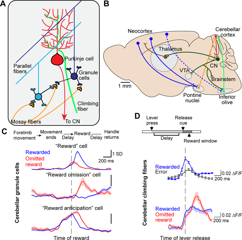
Figure 1. Cerebellar circuits and reward signals.
A. Cerebellar cortex microcircuit. Input arrives via the mossy fiber pathway from specific nuclei in the pons, medulla, and spinal cord, as well as from the climbing fiber pathway from the inferior olive. Each mossy fiber synapses onto ~50 granule cells, and each granule cell receives input from about four mossy fibers. ~100,000 granule cell parallel fiber axons synapse onto each Purkinje cell, while each Purkinje cell receives input from only one climbing fiber. For simplicity, GABAergic interneurons in the cerebellar cortex are omitted from this schematic. Purkinje cells project axons to the cerebellar nuclei (CN), which also receive collaterals from both mossy fibers and climbing fibers (not shown).
B. Connections between the cerebellum and other brain regions. Nearly all subcortically-projecting layer 5 pyramidal neurons throughout the neocortex send an axon collateral to the pontine nuclei. The cerebellum receives mossy fibers from all pontine nuclei neurons, and climbing fibers from inferior olive neurons. The cerebellar nuclei (CN), targets of Purkinje cell axons, project to numerous targets including the cortex via the thalamus, the ventral tegmental area (VTA), and the brainstem nuclei including the inferior olive. Dashed arrows represent indirect input from the cortex to the inferior olive.
C. Reward expectation signaling in cerebellar granule cells. Top, mice executed a forelimb operant task for water reward during two-photon Ca2+ imaging [38]. Bottom, three example granule cell activity profiles. Traces show fluorescence aligned to reward delivery (dashed vertical line) and averaged across either rewarded trials or trials on which reward was unexpectedly omitted. From top to bottom, these three cells were active preferentially during reward delivery, reward omission, or the delay while the mouse waited for the reward. Note that the “reward anticipation” cell remained active longer while the mouse continued waiting following unexpected reward omission, until the mouse gave up and ceased licking (not shown).
D. Reward expectation signaling in cerebellar climbing fibers. Top, water-restricted mice executed a press-and-hold forelimb lever task for water reward during two-photon Ca2+ imaging of Purkinje dendrites [52], whose fluorescence reports climbing fiber activity, in lobule simplex of the cerebellum. Bottom, example climbing fiber activity. Climbing fibers became active preferentially at the time of the lever release on correctly-timed trials, which predicted subsequent reward, but not on error trials, which did not yield subsequent reward. On correctly-timed trials on which the subsequent reward was omitted, a second climbing fiber response was elicited.
C reproduced with permission from [38], D reproduced with permission from [52].

