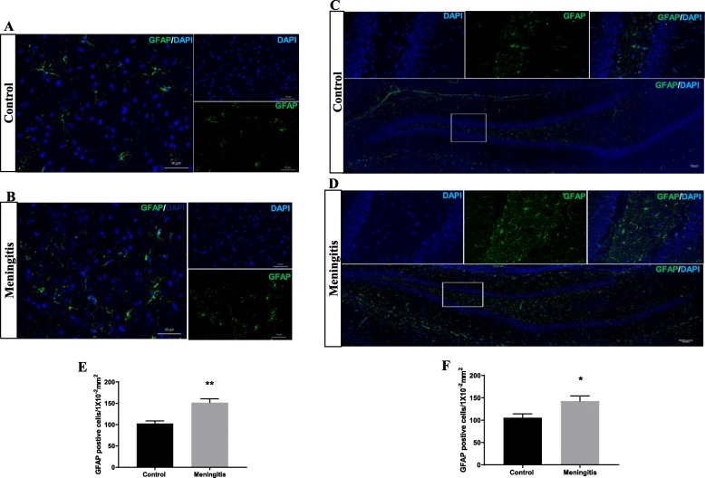
Fig. 8.
Increased astroglial activation 10 days after experimental meningitis induction. Immunofluorescence analysis for GFAP-positive cells in experimental meningitis rats and control rats. a, b Representative microscopic field images (magnification, × 400) from the 10-day group immunostained with GFAP antibodies in the PFC. c, d Representative microscopic field images (magnification, × 200) from the 10-day group immunostained with GFAP in the hippocampus. The number of GFAP-positive cells/1 × 10−2 mm2 in e PFC and f hippocampus in the 10-day group. The results are expressed as the mean ± SEM for n = 4–5 rats. *P < 0.05, **P < 0.01 compared to controls

