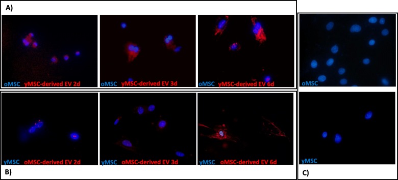
Fig. 3.
Internalisation of MSC-derived EVs as observed by fluorescence microscopy. Images from fluorescence microscopy (magnification, × 40) of the nuclei of MSCs stained with 4′,6-diamidino-2-phenylindole (DAPI) and MSC-derived EVs stained with DiI. a Old MSCs cultured with young MSC-derived EVs at 2, 3 and 6 days. b Young MSCs cultured with old MSC-derived EVs at 2, 3 and 6 days. c Control of young MSCs and old MSCs without added EVs. One representative experiment is shown. oMSC, MSCs from the old group; yMSC, MSCs from the young group; oEV, MSC-derived EVs from the old group; yEV, MSC-derived EVs from the young group

