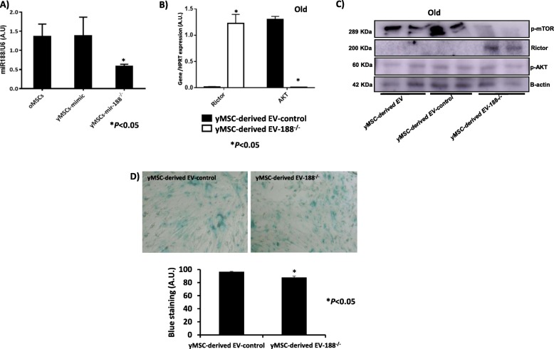
Fig. 7.
Effect of inhibition of miR-188-3p in MSCs. a miR-188-3p expression in old MSCs and young MSCs transfected with mimic or miR-188-3p inhibitor, as determined by RT-qPCR analysis normalised with the expression of U6 small nuclear RNA. b Rictor and AKT gene expressions using RT-qPCR analysis normalised with the expression of HPRT in old MSCs treated with young MSC-derived EVs mimic used as a control and young MSC-derived EVs miR188−/−. *P < 0.05 compared with control was considered statistically significant using Mann-Whitney U and Kruskal-Wallis tests. c Rictor and phosphor-AKT levels in old MSCs treated with young MSC-derived EV mimic used as control and young MSC-derived EVs miR188−/−, using western blot analysis normalised by the expression of β-actin. d Representative images (magnification, × 20) of senescence-associated beta-galactosidase (SA-β-gal) staining of old MSCs with young MSC-derived EVs and with young MSC-derived EVs modified with miR-188-3p inhibitor added to the culture media, showing a higher number of negative cells in young MSC-derived EVs miR-188−/−-treated cells in the densitometry analysis of the staining signal (bottom). Control, MSCs cultured with growth medium without EVs

