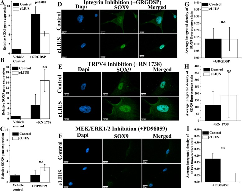
Fig. 3.
cLIUS-induced SOX9 regulation under integrin, TRPV4, and MEK/ERK1/2 inhibition. SOX9 gene expression in MSCs under a integrin inhibition by GRGDSP, b TRPV4 inhibition by RN 1738 (n = 3), and c MEK/ERK1/2 inhibition by PD98059 is shown. Serum-starved 2D cultures of MSCs were treated with inhibitors: 100 μg/ml GRGDSP (integrin inhibitor) or 30 μM RN1738 (TRPV4 inhibitor) for 4 h. Total RNA was collected 1 h after cLIUS stimulation at 14 kPa (5 MHz, 2.5 Vpp) for 5 min and the gene expression of SOX9 was quantified by qRT-PCR. Non-cLIUS-stimulated MSCs incubated in DMSO served as vehicle controls (n = 3). Data represented as a mean ± standard deviation. d–f MSCs were grown in coverslips (n = 4–6 per treatment condition) at an initial seeding density of 1 × 10⁵ cells/well and were treated with inhibitors followed by the cLIUS application at 14 kPa (5 MHz, 2.5 Vpp) for 5 min and fixed in 4% PFA. Confocal micrographs (× 60 magnification) of immunofluorescent staining of SOX9 (green) shows the localization of SOX9 in the MSCs under d integrin inhibition by GRGDSP, e TRPV4 inhibition by RN1738, and f MEK/ERK1/2 inhibition by PD98059. The nucleus was stained with Dapi (blue). Scale bar represents 20 μm. g–i Quantification of SOX9 immunofluorescence intensity in control and cLIUS samples in the presence or absence of inhibitors by ImageJ (n = 10–20). Data are shown as the mean ± standard deviation of samples in triplicate. The p value represents the statistical significance, and n.s represents the non-significant difference as analyzed by Welch’s t-test

