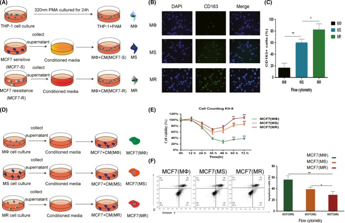
Figure 1.

Crosstalk between tumor‐associated macrophages (TAM) and MCF7 cells. A, Procedure used to obtain macrophages in different microenvironments (MΦ, TAM, TAM from a tamoxifen‐sensitive tumor microenvironment (TME) [MS], and TAM from tamoxifen‐resistant TME [MR]). B, CD163 immunofluorescence (IF) staining in MΦ, MS, and MR macrophages. Single channels and merged images are shown. C, CD163 expression of MΦ, MS, and MR macrophages was analyzed by flow cytometry. D, Procedure used to elucidate the effects of conditioned medium (CM) from different macrophages on MCF7 cells. E, Relative viability of MCF7 (MΦ), MCF7 (MS), and MCF7 (MR) cells treated with 5 μmol/L tamoxifen. F, Apoptosis of MCF7 (MΦ), MCF7 (MS), and MCF7 (MR) cells was analyzed by flow cytometry after adding 5 μmol/L tamoxifen for 24 h. *P < .05, **P < .01
