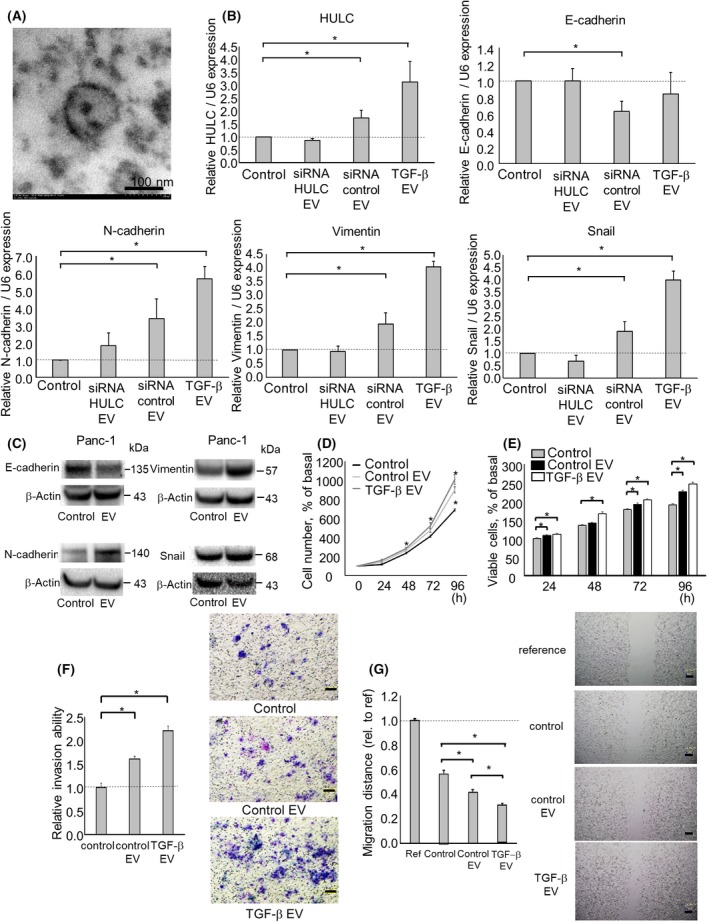
Figure 4.

Intercellular HULC transfer by extracellular vesicles (EVs) during the epithelial‐mesenchymal transition and the phenotype of pancreatic ductal adenocarcinoma cells. Panc‐1 cells (1 × 10⁶ per 10 cm dish) were cultured for 72 h, and EVs were isolated from the conditioned medium by ultracentrifugation. A, Transmission electron micrograph of EVs from Panc‐1 cells. A homogeneous population of particles was obtained. B‐G, EVs were isolated from Panc‐1 cells after transfection with an siRNA to HULC 1 or a control siRNA, or incubated with 10 ug/mL transforming growth factor (TGF‐β for 72 h. Then, EVs were added to recipient Panc‐1 cells. B, RNA was isolated from recipient cells and HULC, E‐cadherin, N‐cadherin, vimentin, and Snail expression was assayed by quantitative RT‐PCR. RNA expression was normalized to that of RNU6B and expressed relative to the value of the control. C, Protein was isolated from recipient Panc‐1 cells and the E‐cadherin, N‐cadherin, vimentin, and Snail levels were analyzed by western blotting. Protein levels were normalized to that of β‐actin. D,E, After 24, 48, 72, and 96 h, cell proliferation was examined by cell counting using Trypan blue (D) and cell viability was examined by MTS assay (E). F, After 24 h, cell invasion was assessed by Transwell assay under an inverted microscope. G, After 24 h, cell migration was assessed by wound healing assay. Bars are means ± SEM of 3 independent experiments. *P < .05. ref, reference; rel., relative
