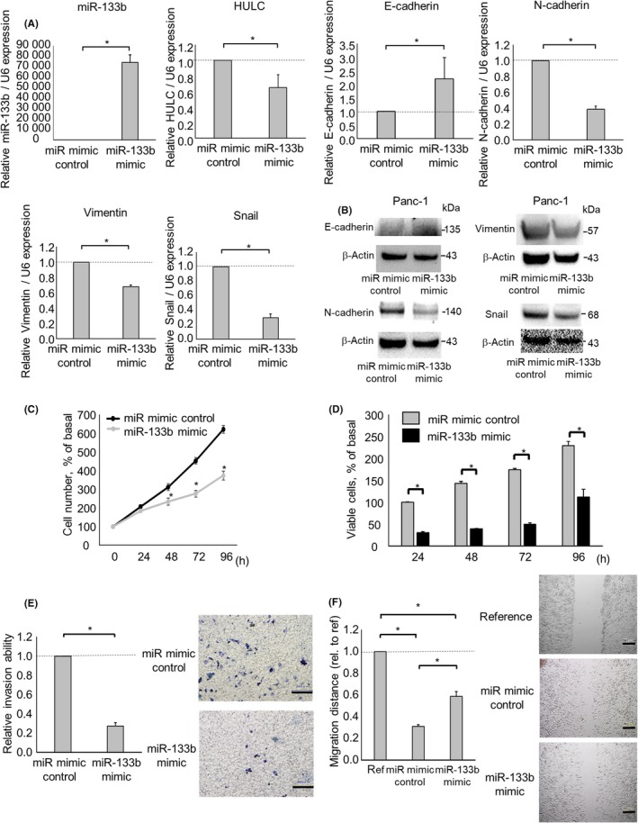
Figure 6.

Effect of microRNA (miR)‐133b overexpression on the epithelial‐mesenchymal transition and phenotype of pancreatic ductal adenocarcinoma cells. Panc‐1 cells were transfected with 12.5 nmol/L miR‐133b or the control mimic. A, After 48 h, RNA was isolated and quantitative RT‐PCR for miR‐133b, HULC, E‐cadherin, N‐cadherin, vimentin, and Snail was carried out. B, After 72 h, protein was extracted, and western blotting was undertaken using Abs against E‐cadherin, N‐cadherin, vimentin, and Snail; protein levels normalized to that of β‐actin are shown. C,D, After 24, 48, 72, and 96 h, cell proliferation was examined by cell counting using Trypan blue (C) and cell viability was examined by MTS assay (D). E, After 24 h, cell invasion was assessed by Transwell assay under an inverted microscope. F, After 24 h, cell migration was assessed by wound healing assay. Bars are means ± SEM of 3 independent experiments. *P < .05. ref, reference; rel., relative
